As Featured In
Industry leader in Detox since 2000
Free Shipping on all US Ground orders
100% Satisfaction or your money back
COMPARE OUR CLEANSES
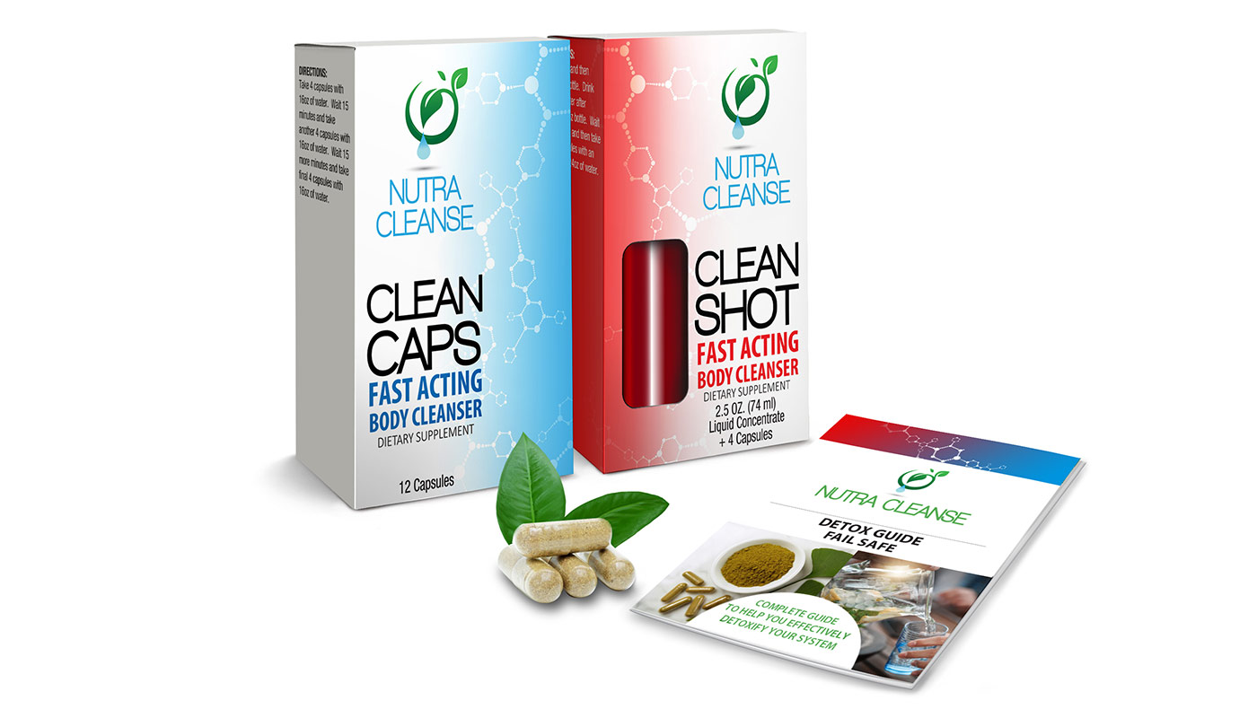
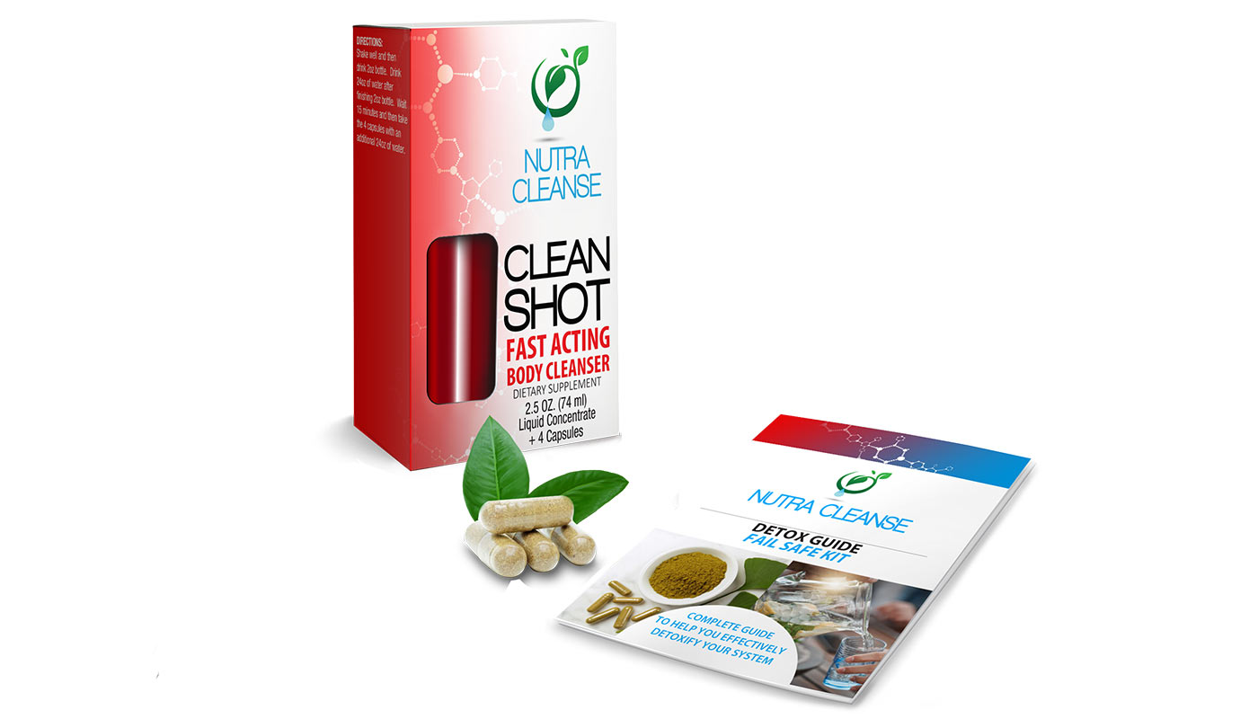
 Same Day Cleanse
$50 - $90
$50 - $90
Urine
Urine
Need Same-Day Results
Need Same-Day Results
Clean for Up to 6 Hours
Clean for Up to 6 Hours
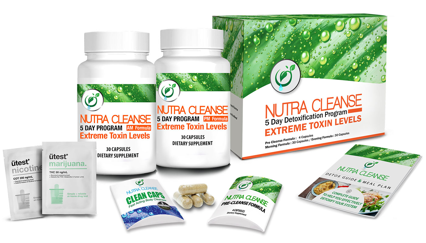
 Permanent Cleanse
$110 - $150
$110 - $150
Urine, BloodUrine, Blood
Have 5 or 10 Days Before Test
Have 5 or 10 Days Before Test
Permanently Clean
Permanently Clean
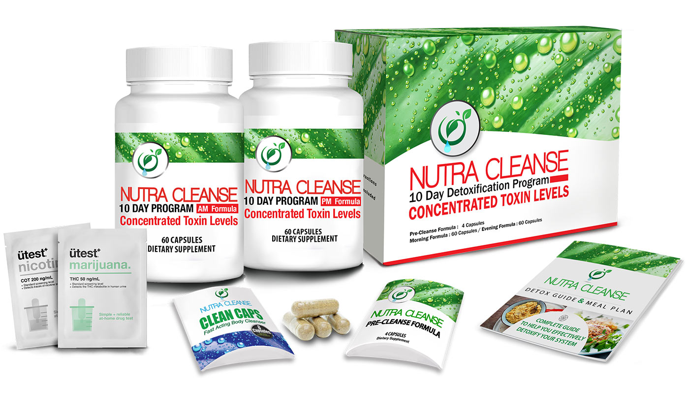
 Total Body Cleanse
$180 - $220
$180 - $220
Urine, Blood, Hair
Urine, Blood, Hair
Have 5 or 10 Days Before TestHave 5 or 10 Days Before Test
Permanently Clean
Permanently Clean
 Cleansing Shampoo
$100 - $220
$100 - $220
Hair
Hair
Need Same-Day Results
Need Same-Day Results
Clean for Up to
24 Hours
Clean for Up to
24 Hours


*Prices have been rounded to the nearest dollar
FREE SHIPPING available on all US Ground orders!
Need it faster? Select Next Day Air at checkout.
See our Shipping Policy for more information.
 Secure Checkout
- 100% Guarantee
- Credit Cards Accepted
- Made in the USA
- GMP Certified
FREE SHIPPING available on all US Ground orders!
Need it faster? Select Next Day Air at checkout.
See our Shipping Policy for more information.
- Secure Checkout
- 100% Guarantee
- Credit Cards Accepted
- Made in the USA
- GMP Certified
FREE SHIPPING available on all US Ground orders!
Need it faster? Select Next Day Air at checkout.
See our Shipping Policy for more information.
- Secure Checkout
- 100% Guarantee
- Credit Cards Accepted
- Made in the USA
- GMP Certified
FREE SHIPPING available on all US Ground orders!
Need it faster? Select Next Day Air at checkout.
See our Shipping Policy for more information.
- Secure Checkout
- 100% Guarantee
- Credit Cards Accepted
- Made in the USA
- GMP Certified